
- Home
- Brain surgery in Tijuana
Brain surgery in Tijuana
Tumours, aneurysms, haemorrhage, hydrocephalus and head trauma. Brain surgery begins long before the operating room: in the careful reading of your imaging.
What brain surgery is, and when it is indicated
Brain surgery covers the procedures that treat lesions inside the skull: tumours, blood collections, vascular malformations, infections and the consequences of head trauma. They share an access route — usually the craniotomy — but very little else: each indication has its own work-up, technique and prognosis.
A surgical indication arises when three things agree: the patient's symptoms, the neurological examination and what the imaging shows. When those three point to the same place, there is a surgical case. When they do not, the prudent course is more investigation, not earlier surgery.
The commonest conditions
The pictures that most often reach this office are brain tumours — gliomas, meningiomas, metastases and pituitary adenomas — head trauma with intracranial haematomas, intracranial aneurysms and subarachnoid haemorrhage, hydrocephalus in children and adults, and ischaemic stroke with mass effect.
Also treated are trigeminal neuralgia, hemifacial spasm, facial palsy, epilepsy with a structural lesion, arachnoid cyst, brain abscess and congenital malformations of the nervous system. Each has its own page in the conditions section.
How an intracranial lesion is investigated
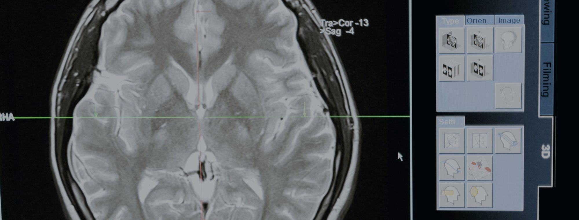
Contrast-enhanced MRI is the reference study: it defines margins, oedema, the relationship to eloquent areas and structural shift. CT is faster and is what emergency departments use, and what shows bone and acute bleeding, but it does not replace MRI.
Where vascular pathology is suspected, CT angiography or cerebral angiography is added. And for lesions near language or motor cortex, planning includes choosing the approach corridor that crosses the least healthy tissue. Surgery starts on the screen, not on the skin.
What happens in theatre, and what is protected
The approach is chosen as a corridor: pterional, subtemporal, retrosigmoid, orbitozygomatic or transsphenoidal, according to where the lesion sits and what lies on the way. The operating microscope, the ultrasonic aspirator and fine bipolar coagulation allow work in a plane of millimetres.
The limit of resection is set by function, not by ambition. When removing the last millimetre of tumour would cost speech, the strength of a hand or vision, a safe resection plus follow-up or radiosurgery is preferred. That decision is discussed beforehand, not improvised inside.
What brain surgery covers
Craniotomy
The access route for almost all intracranial surgery; the bone goes back at the end.
Tumour microsurgery
Resection of gliomas, metastases and other tumours under the operating microscope.
Vascular surgery
Aneurysm clipping and resection of arteriovenous malformations.
Haematoma evacuation
Epidural and subdural haematoma, acute traumatic or chronic in older adults.
Shunting for hydrocephalus
A valved system diverting cerebrospinal fluid to the abdomen.
Skull base surgery
Approaches designed to pass between cranial nerves and major vessels.
Functional neurosurgery
Trigeminal neuralgia, hemifacial spasm and lesional epilepsy.
Head trauma
Depressed fractures, contusions and decompressive craniectomy.
Bring your imaging and settle the question
One consultation with your MRI or CT in hand is worth more than ten internet searches. Book by phone or through the form and you will be told what is there, what it means and what your options are.