
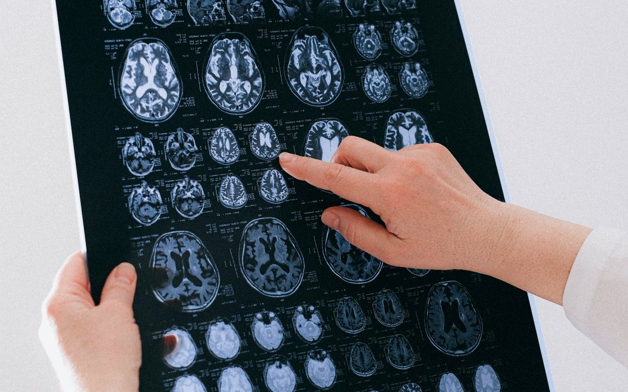
Brain tumour microsurgery
Removal of gliomas, metastases and other brain tumours under the microscope, with neurological function as the limit of the surgical gesture.
See the procedure
Not every brain tumour is cancer, and not all are operated the same way. The first task is to know which type it is, where it sits and what it is doing to the brain.
An intracranial tumour can arise from nervous tissue itself, from the meninges that wrap it, from the pituitary or from the cranial nerves; or it can arrive from another organ as a metastasis. That distinction is the first one made in consultation, because it changes prognosis, treatment and how urgently one must act.
Symptoms depend less on tumour type than on location and growth rate. A slow-growing tumour in a silent area can reach a considerable size before giving any sign; a small one beside the motor cortex may announce itself with weakness of one hand within weeks. The same diagnosis therefore produces very different stories.
The work-up begins with contrast-enhanced MRI, the reference study. CT is useful in emergencies and for bone and bleeding, but it does not replace MRI. The definitive diagnosis, however, comes from histopathology of the tissue: until then we work with probabilities, not certainties.
The procedures that may be indicated for this condition. Which one applies to you — if any does — is decided on your imaging and your examination.
Removal of gliomas, metastases and other brain tumours under the microscope, with neurological function as the limit of the surgical gesture.
See the procedure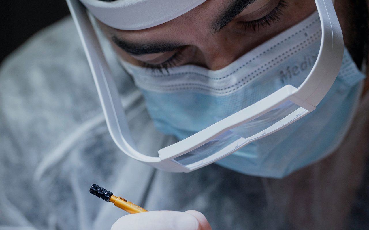
A temporary bone window to reach the brain. The bone is replaced and fixed at the end — no gap is left behind.
See the procedure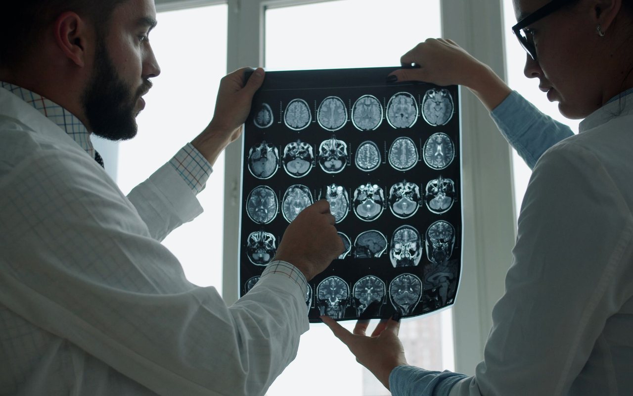
Resection of convexity, falcine, sphenoid wing and skull base meningiomas, with early devascularisation.
See the procedure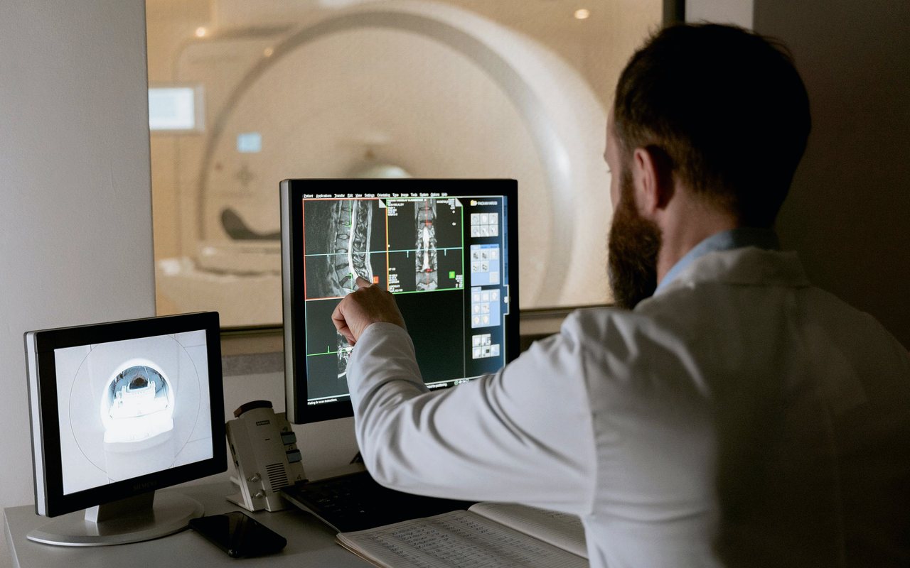
Resection of pituitary adenomas, with endocrine assessment and visual field testing before and after surgery.
See the procedureLoss of consciousness, the worst headache of your life, sudden weakness on one side, difficulty speaking, a seizure, projectile vomiting, or loss of bladder control together with back pain. Go to an emergency department immediately and call (664) 104-9153.
It is very unlikely. The overwhelming majority of headaches are migraine or tension-type, not tumours. What is concerning is a headache that is new and different from your usual one, wakes you at night, worsens on coughing or bending, or comes with vomiting, seizure, double vision or weakness. That combination does warrant imaging.
The great majority are not. Rare genetic syndromes such as neurofibromatosis or Li-Fraumeni raise the risk, but they account for a small fraction of cases.
Large epidemiological studies have not demonstrated that link. Research continues, but there is no solid evidence supporting that cause today.
It depends on the tumour. A small meningioma can be watched for years; a tumour with oedema and mass effect cannot. At your visit you are told explicitly whether your case allows time for a second opinion or whether action is needed soon.
One consultation with your MRI or CT in hand is worth more than ten internet searches. Book by phone or through the form and you will be told what is there, what it means and what your options are.