
- Home
- Procedures
- Brain tumour surgery in Tijuana
Brain tumour surgery in Tijuana
Resection of brain tumours under the operating microscope, aiming for maximum tumour with minimum deficit. Strategy changes with tumour type, location and what surrounds it.
Not every brain tumour is operated the same way
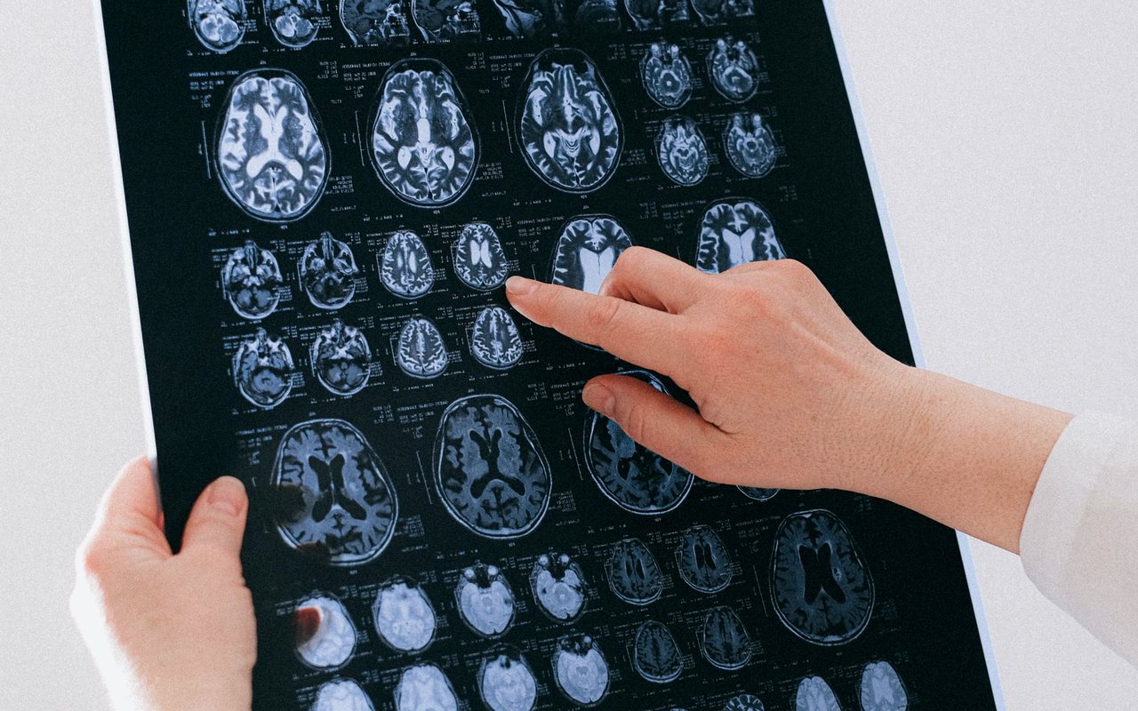
“Brain tumour” is an enormous category. A convexity meningioma, a single lung metastasis, a diffuse glioma that weaves between the fibres of the brain and a pineal region tumour share neither technique, nor prognosis, nor even the decision to operate. That is why the first consultation is spent on the contrast MRI, defining what kind of lesion we are looking at and what surgery would actually gain.
In some cases the goal is complete resection, which can be curative. In others the goal is tissue for diagnosis and volume reduction to lower intracranial pressure, while radiation and medical oncology do the rest. Confusing those two goals is what produces expectations that later go unmet.
Function sets the limit, not the instrument
In brain tumour surgery the question is not only how much can be removed, but how much can be removed without leaving the patient worse than they arrived. Near the motor area, the language cortex or the visual pathways, resection stops where the tissue that supports a function you use every day begins.
Working within that margin calls for the operating microscope, the ultrasonic aspirator and fine bipolar coagulation, with planning done beforehand on the MRI. The specimen goes to histopathology, because the tumour's definitive name comes from the pathologist, not from the image.
What the procedure involves
When it is indicated
- Low- and high-grade glioma with a surgical indication
- Single or accessible brain metastasis with the primary disease under control
- Tumour with mass effect and raised intracranial pressure
- Lesion needing histological diagnosis before treatment can be decided
- Intraventricular tumour obstructing cerebrospinal fluid circulation
- Tumour recurrence with an indication for repeat resection
Techniques and resources
- Operating microscope
- Ultrasonic aspirator
- Bipolar coagulation
- Contrast MRI
- Histopathology
- Neuronavigation where available
Frequently asked questions about brain tumour microsurgery
No. Many intracranial tumours are benign — meningiomas, pituitary adenomas, schwannomas — and grow slowly without invading. Others are malignant. Behaviour is defined by histopathology of the specimen, not by the fright of the first day.
It depends on type and location. Well-defined tumours away from critical areas are usually resected completely. Diffuse gliomas infiltrate healthy tissue, and chasing one hundred per cent at any cost there can cost a function. That is discussed before the operation, not after.
In some cases yes, decided once pathology is back. Surgery, radiotherapy and medical treatment are parts of one plan; which parts apply to you depends on type, grade and how much was resected.
Bring your imaging and settle the question
One consultation with your MRI or CT in hand is worth more than ten internet searches. Book by phone or through the form and you will be told what is there, what it means and what your options are.