
- Home
- Procedures
- Lumbar microdiscectomy in Tijuana
Lumbar microdiscectomy in Tijuana
Removal of the disc fragment compressing a nerve root, through a small incision and under the microscope. It is the operation that takes away sciatica — not back pain.
What it solves, and what it does not
It is worth being exact here, because this is where most disappointment is created. Microdiscectomy is designed to remove compression from a nerve root, and what it relieves fastest and most consistently is the pain running down the leg: sciatica. Background lumbar pain, the ache across the belt line, improves less and sometimes not at all.
So the best candidate is someone with clearly radicular pain, a herniation on MRI matching exactly the level and side of their symptoms, who has already had six to twelve weeks of properly delivered conservative treatment without result. When those three line up, results are usually very good.
The technique and what follows
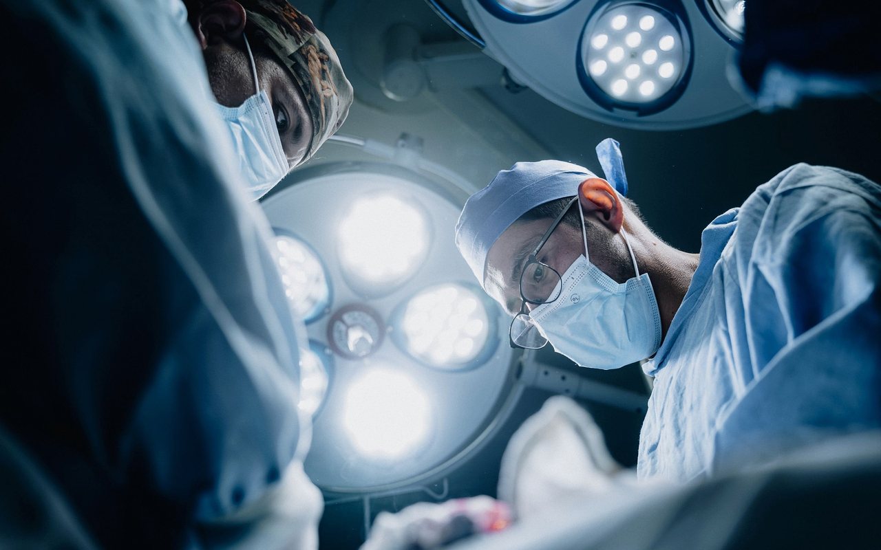
The incision measures around three centimetres over the midline. Muscle is retracted rather than cut, a small portion of ligament or lamina is removed to see the root, and under the microscope the herniated fragment is identified and extracted. The root is left free and mobile before closure. The whole disc is not removed — only the fragment that compresses.
Most patients walk the same day and go home within 24 hours. Return to desk work usually falls between the second and fourth week; heavy work, later. Rehabilitation afterwards is not optional: strengthening the trunk is what lowers the risk of a new herniation.
What the procedure involves
When it is indicated
- Lumbar disc herniation with sciatica unresponsive to conservative care
- Motor deficit from root compression (foot drop, weakness climbing stairs)
- Disabling radicular pain with exact clinical-radiological correlation
- Extruded or migrated herniation with frank root compression
- Cauda equina syndrome, as an immediate emergency
- Recurrent herniation at the same level with radicular symptoms
Techniques and resources
- Operating microscope
- Tubular retractor or blade
- High-speed drill
- Lumbar MRI
- Microdiscectomy instruments
- Fluoroscopy for level marking
Frequently asked questions about lumbar microdiscectomy
It mainly takes away the pain running down the leg. Lumbar pain may improve, but that is not what this operation is designed to treat. If your main complaint is the low back rather than the leg, whether the herniation really is the cause needs reviewing before surgery.
Driving, generally within a few days, once you are off strong analgesics. Desk work between the second and fourth week. Physical work or lifting, according to progress, and normally not before six weeks.
Yes, recurrence occurs in a minority of cases, more often in smokers, in people carrying excess weight, and in those who return early to lifting. Trunk strengthening, stopping smoking and careful lifting technique are the three measures that help most.
Bring your imaging and settle the question
One consultation with your MRI or CT in hand is worth more than ten internet searches. Book by phone or through the form and you will be told what is there, what it means and what your options are.